
A Rare Case of Extensive Bilateral Fibroadenomas in a Young Woman
Keywords:
Fibroadenoma, Breast, Benign, Doppler Ultrasound, Radiology, breastfeedingAbstract
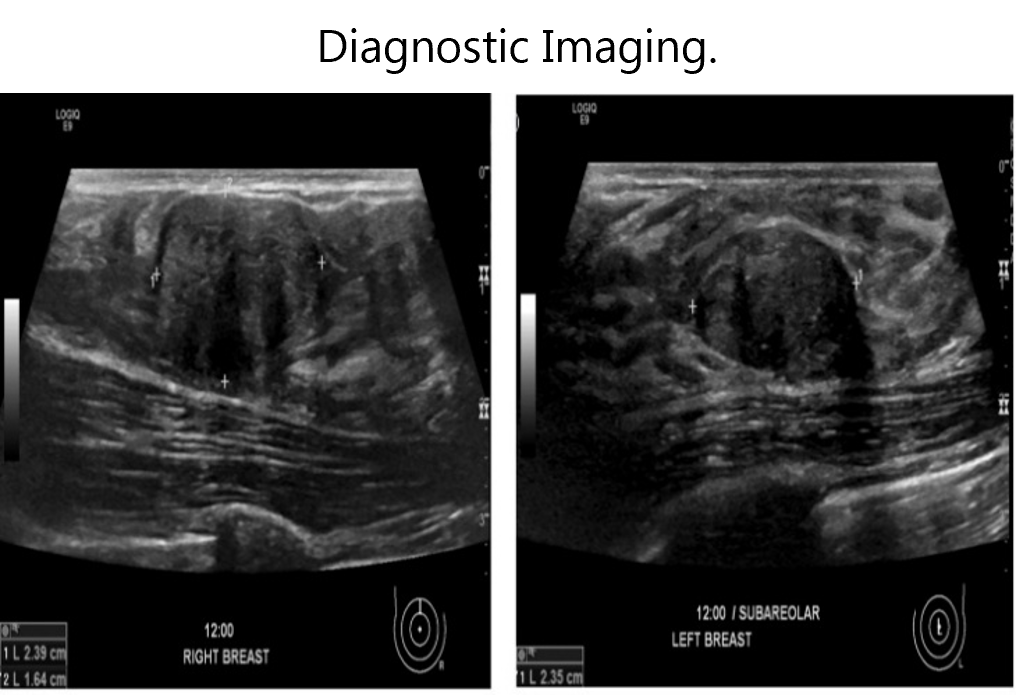
Background Fibroadenomas are benign, solid nodules in the breast commonly found in women less than 35 years of age. Typically, fibroadenomas do not exceed 3-4 per breast, and the occurrence of five or more is uncommon. Case We present a case of an 18-year-old female, initially diagnosed with bilateral fibroadenomas, progressing to an extensive presentation of over 25 fibroadenomas per breast by age 26, a rarity in the medical literature. Despite recommendations for a mastectomy due to the lesion burden, the patient, after consulting with the physician and understanding the risk of progression, opted for watchful waiting, citing concerns about future breastfeeding. Conclusion This case underscores the importance of shared decision-making in managing extensive fibroadenomas in young women, considering their impact on future reproductive choices. We aim to raise awareness of such uncommon presentations and encourage further reporting to broaden understanding and management strategies.
References
Salati SA. Breast fibroadenomas: a review in the light of current literature. Pol Przegl Chir. 2020;93(1):40–8.
Santen RJ, Mansel R. Benign breast disorders. N Engl J Med. 2005;353(3):275–85.
Nassar A, Visscher DW, Degnim AC, Frank RD, Vierkant RA, Frost M, et al. Complex fibroadenoma and breast cancer risk: a Mayo Clinic Benign Breast Disease Cohort Study. Breast Cancer Res Treat. 2015;153(2):397–405.
Román M, Louro J, Posso M, Vidal C, Bargalló X, Vázquez I, et al. Long-term risk of breast cancer after diagnosis of benign breast disease by screening mammography. Int J Environ Res Public Health. 2022;19(5):2625.
Williamson ME, Lyons K, Hughes LE. Multiple fibroadenomas of the breast: a problem of uncertain incidence and management. Br J Surg. 1993;75(3):161–3.
Balakrishnan A, Yang J, Beattie CW, Gupta TKD, Nandi S. Estrogen receptor in dissociated and cultured human breast fibroadenoma epithelial cells. Cancer Lett. 1987;34(3):233–42.
Ramala SR, Chandak S, Chandak MS, Annareddy S. A comprehensive review of breast fibroadenoma: correlating clinical and pathological findings. Cureus. 2023;15(12):e49948.
Im CJ, Miller A, Balassanian R, Mukhtar RA. Early onset, multiple, bilateral fibroadenomas of the breast: a case report. BMC Womens Health. 2021;21(1):178.
Panda SK, Patro B, Mishra J, Dora RK, Subudhi BSK. Multiple fibroadenomas in bilateral breasts of a 46-year-old Indian woman – a case report. Int J Surg Case Rep. 2014;5(5):262–4.
Zhang RR, Bevan S, Sun P, Lu JZ, Peng Y. Unusual presentation of multiple fibroadenomas in bilateral breasts and axillary accessory breast. Breast Cancer (Auckl). 2012;6:13–6.
Shibata J, Fujii M, Kato T. Multiple fibroadenomas of the breasts: a cause of monthly chest pain. Postgrad Med J. 2022;98(1166):e28.
Aga MB, Mishra AA, G. Multiple bilateral giant juvenile fibroadenomas of breast. Eur J Surg. 2000;166(10):828–30
Published
How to Cite
License
Copyright (c) 2025 Cory Dixon, Sawyer Longley, Aaron Tillman, Britton Ethridge, Samuel Armstrong

This work is licensed under a Creative Commons Attribution 4.0 International License.
Authors who publish with this journal agree to the following terms:
- The Author retains copyright in the Work, where the term “Work” shall include all digital objects that may result in subsequent electronic publication or distribution.
- Upon acceptance of the Work, the author shall grant to the Publisher the right of first publication of the Work.
- The Author shall grant to the Publisher and its agents the nonexclusive perpetual right and license to publish, archive, and make accessible the Work in whole or in part in all forms of media now or hereafter known under a Creative Commons Attribution 4.0 International License or its equivalent, which, for the avoidance of doubt, allows others to copy, distribute, and transmit the Work under the following conditions:
- Attribution—other users must attribute the Work in the manner specified by the author as indicated on the journal Web site; with the understanding that the above condition can be waived with permission from the Author and that where the Work or any of its elements is in the public domain under applicable law, that status is in no way affected by the license.
- The Author is able to enter into separate, additional contractual arrangements for the nonexclusive distribution of the journal's published version of the Work (e.g., post it to an institutional repository or publish it in a book), as long as there is provided in the document an acknowledgment of its initial publication in this journal.
- Authors are permitted and encouraged to post online a prepublication manuscript (but not the Publisher’s final formatted PDF version of the Work) in institutional repositories or on their Websites prior to and during the submission process, as it can lead to productive exchanges, as well as earlier and greater citation of published work. Any such posting made before acceptance and publication of the Work shall be updated upon publication to include a reference to the Publisher-assigned DOI (Digital Object Identifier) and a link to the online abstract for the final published Work in the Journal.
- Upon Publisher’s request, the Author agrees to furnish promptly to Publisher, at the Author’s own expense, written evidence of the permissions, licenses, and consents for use of third-party material included within the Work, except as determined by Publisher to be covered by the principles of Fair Use.
- The Author represents and warrants that:
- the Work is the Author’s original work;
- the Author has not transferred, and will not transfer, exclusive rights in the Work to any third party;
- the Work is not pending review or under consideration by another publisher;
- the Work has not previously been published;
- the Work contains no misrepresentation or infringement of the Work or property of other authors or third parties; and
- the Work contains no libel, invasion of privacy, or other unlawful matter.
- The Author agrees to indemnify and hold Publisher harmless from the Author’s breach of the representations and warranties contained in Paragraph 6 above, as well as any claim or proceeding relating to Publisher’s use and publication of any content contained in the Work, including third-party content.
Enforcement of copyright
The IJMS takes the protection of copyright very seriously.
If the IJMS discovers that you have used its copyright materials in contravention of the license above, the IJMS may bring legal proceedings against you seeking reparation and an injunction to stop you using those materials. You could also be ordered to pay legal costs.
If you become aware of any use of the IJMS' copyright materials that contravenes or may contravene the license above, please report this by email to contact@ijms.org
Infringing material
If you become aware of any material on the website that you believe infringes your or any other person's copyright, please report this by email to contact@ijms.org


