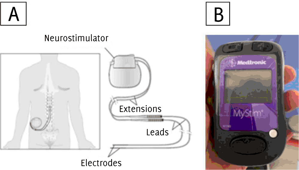
A: Implanted Parts of an Internal Neurostimulator System. B: External Programming Unit of the MyStim Spinal Cord Neurostimulator Model 97740
Shyla Gupta1, Cathy Shaw, Sohaib Haseeb, Adrian Baranchuk
doi: http://dx.doi.org/10.5195/ijms.2020.614
Volume 8, Number 2: 145-147
Received 18 06 2020: Rev-request 04 07 2020: Rev-recd 06 07 2020: Accepted 17 08 2020
ABSTRACT
Background:Neurostimulator devices produce electrical oscillations that may prevent accurate diagnosis of an ECG.
The Case:We present the case of a 68-year-old man who came to the emergency department with chest pain and a spinal cord neuromodulator device in situ to treat his polymyalgia rheumatica. A 12-lead ECG was obtained to determine the cause of the chest pain, and atrial fibrillation was wrongly diagnosed.
Conclusion:This case reiterates the value of recognizing this uncommonly encountered ECG artifact to avoid unnecessary mistakes in interpretation of heart rhythms.
Keywords: Neurostimulator devices; ECG artifact; Atrial fibrillation (Source: MeSH-NLM).
Acquisition of high-quality surface 12-lead electrocardiograms (ECGs) in the emergency department (ED) is paramount to facilitate interpretation of different clinical presentations.1 Inability to obtain adequate recordings significantly diminish the capacity of the interpreter, leading to potentially serious medical errors.
We present the case of a 68-year-old male with a history of polymyalgia rheumatica with pain refractory to usual care and implanted with a neuromodulation device (MyStim Neuromodulator Device, Medtronic) (Figure 1A). The patient reported a history of metastatic lung cancer, for which he underwent surgical removal of a lung mass via thoracotomy. On this occasion, he presented to the ED with complaints of chest pain. A 12-lead ECG was obtained; however, the automatic analysis of the ECG machine was unable to determine whether the patient had a pacemaker. From this ECG, there was an erroneous diagnosis of atrial fibrillation (Figure 2A). The patient had an external programmer (MyStim Programmer Model 97740, Medtronic) with the ability to inactivate the neurostimulator and adjust the stimulation level (Figure 1B). After the neuromodulation device was switched off, a repeat ECG showing normal sinus rhythm was obtained (Figure 2B).
Figure 1A: Implanted Parts of an Internal Neurostimulator System. B: External Programming Unit of the MyStim Spinal Cord Neurostimulator Model 97740
A: ECG with atrial fibrillation obtained with neuromodulator active. B: ECG without artifact, obtained after turning off the neuromodulator device

Neuromodulation stimulators are inserted in a wide variety of patients in the context of a range of conditions. Devices like these target different anatomical sites, such as the deep brain and spinal cord. Neurostimulator devices produce electrical oscillations that may hinder the procurement of an ECG and can generate artifacts that may interfere with the accurate diagnosis of the data attained.2 The neurostimulator controller is a miniature hand-held, wireless device, similar to a remote controller. It delivers electrical signals to the epidural space near the spine through lead-wires.3 It is used for the treatment of polymyalgia rheumatica chronic pain. Spinal cord stimulation reduces chronic pain and improves the ability to go about daily activities by modifying and masking the pain signal before it reaches the brain.4
Highlights:
The device works by adjusting amplitude, pulse width, and rate of pulses delivered per second, according to the therapy prescribed by the physician.
The stimulation induces artifact in the ECG tracing, posing different difficulties to a precise analysis of the surface ECG. In this case, momentarily impairing the device provided an accurate ECG recording, showing a normal ECG instead of wrongly diagnosing atrial fibrillation (or any perceived arrhythmia). Other common sources of interference, such as Parkinson disease and tremors, hearing aid devices or sacral neuromodulators, may also act as a barrier to accurate electrocardiographic diagnosis.5
Similarly, deep brain stimulation is another form of electrical interference that has been shown to cause ECG artifact. Because the ECG can pick up electrical activity created by these stimulators, this brings to light other potential sources of interference, artifacts are only visible when neurostimulators are in monopolar mode. 6 This is likely because in bipolar mode, the electrode contact in the brain does not possess enough magnitude to create a notable interference.
Another example of interference comes from transcutaneous electrical nerve stimulators (TENS). TENS produce electric currents that interfere with ECG machines.7 Artifact can occur depending on the frequency and amplitude through which skin electrodes are placed. Additionally, artifact can occur if ECG leads are placed incorrectly, and mimic pathology like in this case.8 Proper ECG interpretation depends on several aspects of clinical care.
Recognizing different sources of artifact, learning how to facilitate a situation where ECG artifact is minimized, and being able to collect a proper ECG is quite essential to maintaining high level care.9 Being aware of the effects of different neuromodulator devices is important for both ECG technicians and physicians.10 Rapid recognition of sources of electromagnetic interference improves surface ECG recordings quality facilitating the accurate diagnosis or exclusion of different medical conditions relating to cardiac electrophysiology.11
None
The Authors have no funding, financial relationships or conflicts of interest to disclose.
Conceptualization, Investigation, Methodology, & Validation: SG, AB. Formal Analysis: CS, AB. Project Administration: AB. Resources: SG, CS. Supervision: SG, SH, AB. Visualization: SG, SH. Writing – Original Draft Preparation: SG. Writing – Review & Editing: SG, SH, AB.
1.Rosen AV, Koppikar S, Shaw C, Baranchuk A. Common ECG Lead Placement Errors. Part I: Limb Lead Reversals. Int J Med Students. 2014 Jul-Oct;2 (3):92–8.
2.Suarez-Fuster L, Alexander B, Renaud R, Shaw C, Baranchuk A. Electrocardiographic interference by a sacral neuromodulation device. J Electrocardiol. 2017;50(4):518–519.
3.Medtronic: MedicalExpo.” The Online Medical Device Exhibition, MedicalExpo, https://www.medicalexpo.com/prod/medtronic/product-70691-622641.html.
4.Spine, Mayfield Brain &. “Spinal Cord Stimulation, SCS, Spinal Cord Stimulators.” Mayfieldclinic.com, mayfieldclinic.com/pe-stim.htm.
5.Diez JC, Shaw C, Baranchuk A. Electrocardiogram interference by a neurostimulator. J Electrocardiol 2010, 43:301.
6.Constantoyannis C, Heilbron B, Honey CR. Electrocardiogram artifacts caused by deep brain stimulation. Can J Neurol Sci. 2004;31(3):343–346.
7.Suarez-Fuster L, Oh C, Baranchuk A. Transcutaneous electrical nerve stimulation electromagnetic interference in an implantable loop recorder. J Arrhythm. 2017;34(1):96–97.
8.Rosen AV, Koppikar S, Shaw C, Baranchuk A. Common ECG Lead Placement Errors. Part II: Precordial Misplacements. Int J Med Students. 2014 Jul-Oct;2 (3):99–103.
9.Diez JC, Shaw C, Baranchuk A. Electrocardiogram interference by a neurostimulator. J Electrocardiol. 2010;43 (4):301.
10.García-Niebla J. The Quest for Quality Electrocardiographic Recording. Int J Med Students. 2014 Jul-Oct;2 (3):87–9.
11.Baranchuk A, Kang J, Shaw C, Witjes R. Electromagnetic interference produced by a hearing aid device on ECG recording. J Electrocardiol 2008;41(5):398–400.
Shyla Gupta, 1 Undergraduate student. Queen's University. Kingston Health Sciences Centre, Ontario, Canada
Cathy Shaw, 2 RN. Kingston Health Sciences Centre, Ontario, Canada
Sohaib Haseeb, 3 BSc. Kingston Health Sciences Centre, Ontario, Canada
Adrian Baranchuk, 4 MD, FACC, FRCPC, FCCS. Kingston Health Sciences Centre, Ontario, Canada
Francisco J. Bonilla-Escobar, Editor
Paul MacDaragh Ryan, Student Editor
Andrew Stanton Kucey, Student Editor
About the Author: Shyla Gupta is an undergraduate student at Queen's University, pursuing her bachelor's degree in Health Sciences. Sohaib Haseeb is a 1st year medical student of James Cook University College of Medicine and Dentistry in Townsville, Queensland, Australia.
Correspondence: Adrian Baranchuck, MD, FACC, FRCPC, FCCS, Address: 76 Stuart St, Kingston, ON K7L 2V7, Canada Email: barancha@kgh.kari.net
Cite as: Gupta S, Shaw C, Haseeb S, Baranchuck A. ECG Artifact by a Spinal Cord Neurostimulator: A Case Report. Int J Med Students. 2020 May-Aug;8(1):145-7.
Copyright © 2020 Shyla Gupta, Cathy Shaw, Sohaib Haseeb, Adrian Baranchuk
This work is licensed under a Creative Commons Attribution 4.0 International License.
International Journal of Medical Students, VOLUME 8, NUMBER 2, August 2020