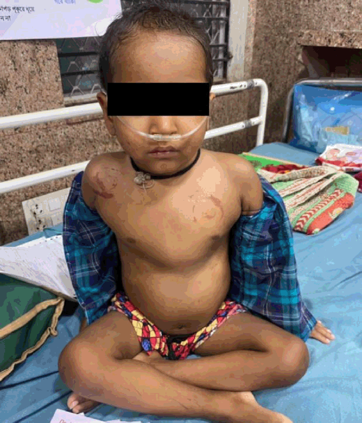
Chest Examination Shows Pectus Carinatum

Legend: The child is orthopneic, he inhales oxygen through nasal prongs. Pectus carinatum also known as pigeon chest, is a bony deformity is which is probably present due to long standing lung infection.
Highlights:Anuva Dasgupta1, Dibyendu Raychaudhuri2
doi: http://dx.doi.org/ijms.2024.2176
Volume 12, Number 4: 468-472
Received 16 07 2023; Rev-request 10 09 2023; Rev-request 29 07 2024; Rev-request 25 10 2024; Rev-recd 16 09 2023; Rev-recd 31 07 2024; Rev-recd 02 11 2024; Accepted 06 11 2024
ABSTRACT
Background:Bronchiectasis is a disorder marked by the destruction of smooth muscle and elastic tissue caused by inflammation, resulting in the permanent expansion of bronchi and bronchioles. It can occur following a single severe episode or repeated episodes of pneumonia, as well as exposure to tuberculosis.
The Case:A child reported with cough and cold for 7 days, with mild fever. He was admitted to the hospital due to breathing difficulties and facial swelling. The clinical exam showed crepitation, wheezing, and pectus carinatum. The patient has a history of multiple hospital admissions due to pneumonia, respiratory distress, and exposure to tuberculosis. His mother was diagnosed and treated for tuberculosis when he was 3 months old. The condition of the patient was evaluated using ultrasonographic examination, chest radiograph, and High-Resolution Computed Tomography of thorax.
Conclusion:High-resolution Computed Tomography (HRCT) scanning is the preferred diagnostic test as it helps to identify the pathologic changes and the exact extent through which it has taken place. Early intervention plays a critical role in reducing severe complications like hemoptysis and cor pulmonale. The current treatment options consist of antibiotics, bronchodilators, anti-inflammatory medications, and physical therapy. The patient was treated using steroids, anti-microbials, and inhalational bronchodilators. Complete symptom resolution was noted in two weeks from date of admission. He also seemed to be doing well in the follow-up visit, one week post discharge. Severe cases may require injectable antibiotics. As a widespread condition in India, early diagnosis and treatment with suitable antimicrobials is critical for a positive outcome.
The principal conditions associated with bronchiectasis are obstruction and infection.1 Infections primarily originate from issues with airway clearance, which cause bronchi and bronchioles to enlarge irreversibly.2 Vertical airways are notably affected, while distal bronchi and bronchioles are more severely affected. The bronchi and bronchioles are typically so dilated that they can be tracked to the pleural surface.3
Clinical signs include a strong, persistent cough, dyspnea, expectoration of foul-smelling, occasionally bloody sputum, and orthopnea in severe cases.1 Upper respiratory tract infections and the introduction of pathogenic organisms causes episodic symptoms. As a result of improved therapeutic methods brain abscesses, amyloidosis, and cor pulmonale are less common complications.2
Histologic findings are influenced by the level of activity and duration of the disease.2 In severe active cases, the lining epithelium of the bronchiolar walls are desquamated. There may be ulceration along with inflammatory exudation. The bronchiolar lumen may be entirely or partially destroyed by peribronchiolar fibrosis, bronchial, and bronchiolar wall fibrosis.3
Treatment for bronchiectasis in India currently only involves managing symptoms, and there is no established protocol. The management of the disease is less than ideal, with over 60% of patients being treated similarly to those with obstructive airway diseases using inhaled corticosteroids, long-acting beta agonists, or both, despite the fact that only 35% of patients show an obstructive pattern on spirometry. More evidence-based treatment, like low-dose macrolides, inhaled antibiotics like tobramycin and colistin, is used in less than 10% of cases.1
Bronchiectasis was thought to be an orphan disease that seldom progressed to severe consequences, especially after the introduction of newer antimicrobials. The incidence of bronchiectasis varies widely, ranging from 67 to 566.1 per 100,000 people in Europe and North America and reaching as high as 1200 per 100,000 individuals aged 40 years or older in China. A comparison of data revealed that Indian patients with bronchiectasis exhibited notable differences from those in the developed world. Patients in India were generally younger (with a mean age of 56 years) and more commonly male (56.9%). Previous tuberculosis was identified as the most frequent underlying cause of bronchiectasis at a rate of 35·5%. Notably, bronchiectasis is emerging as one of the top three chronic airway inflammatory diseases globally, alongside chronic obstructive pulmonary disease and asthma. Understanding the disease burden is imperative for the improvement of the global management of bronchiectasis.1
Patients experience a highly variable clinical course which ranges from complete resolution if diagnosed early to long-term impairment of quality of life and high healthcare costs through exacerbations, hospitalizations, and premature mortality.1 Complications include lung abscess, empyema, atelectasis, cor pulmonale, persistent bacterial bronchitis, and recurrent pleurisy.3
This case is noteworthy because it illustrates bronchiectasis in an Indian child that proceeded to the severe complication of transmediastinal herniation. It is relatively common albeit under-diagnosed in low- and middle-income countries.6
The patient's mother provided written consent for her son's situation to be discussed in a case study.
A 3.5-year-old Indian boy, weighing 11 kgs (who is underweight and falls in the 1st percentile according to WHO weight for age percentile chart),4 presented with a 7 day history of productive cough, cold, and low-grade fever with dyspnea and respiratory distress for past 4 days. Facial swelling is present. He was admitted to the OPD. Patient was apparently well 10 days prior to discharge from the hospital where he was admitted for pneumonia. Child is developmentally normal based on fine motor skills, gross motor skill, language, and social interaction.
The patient's previous medical background was acquired from his mother, revealing that the patient was hospitalized at the age of one month for pneumonia and then at seven months, three years, and four months for symptoms such as coughing, colds, and respiratory distress. He has also visited the local doctor multiple times due to respiratory distress. His mother received a diagnosis of pulmonary tuberculosis (TB) using Cartridge Based Nucleic Acid Amplification Test and was treated with Isoniazid, Rifampicin, Pyrazinamide and Ethambutol when the patient was 3 months old. She discontinued breast-feeding him owing to her tuberculosis diagnosis, and then formula milk was administered. Prenatal history of the patient includes a single mother, born at term, normal vaginal delivery weighing 3.5 kg. Patient has received bacille Calmette-Guerin (BCG) vaccine at birth and 0 and 1st dose of Oral Polio Vaccine (OPV) and Hepatitis B vaccine. He has not received further immunization. He has not received Pentavalent vaccines. This leaves him unprotected from respiratory pathogens such as Corynebacterium diptheriae, Bordetella pertussis, and Haemophilus inflenzae type b. Moreover, BCG vaccine provides immunization only against extra-pulmonary forms of tuberculosis.
Upon examination in a seated position, pectus carinatum is identified by an excessively protruding sternum and the chest having a triangular shape Figure 1. The right side of the chest has a unilateral depression due to bronchiectatic changes and the collapse of the basal segment of the right lung along with decreased movement on the right side of the chest.
Figure 1Chest Examination Shows Pectus Carinatum

Dyspnea is present. On palpation, the patient's chest has an asymmetrical movement. Dullness is heard on percussion of the chest. Vocal fremitus is diminished. Auscultation reveals characteristic breath sounds on left side of the chest but diminished breath sounds on the right side. Rhonchi and localized coarse crepitation are heard which are restricted to the right lung base. S1 and S2 heart sound are prominent. Apex beat palpated at 4th intercostal space towards the left border of the sternum.
The patient has persistent, productive cough without blood. Clubbing of fingers and cyanosis are not seen.
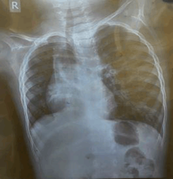
Digital chest radiograph shows bronchiectatic changes. Ultrasonogram (USG) revealed multiple sub-centric, mesenteric lymph nodes, and right-sided mild pleural effusion.
High-Resolution Computed Tomography (HRCT) of thorax revealed collapse of basal segment of right lung, trans-mediastinal space shift of the left upper lobe, and bi-lateral bronchiectatic changes. This indicates the trans-mediastinal herniation of left upper lobe of the lung or the protrusion of the lungs past the mediastinum's anatomic boundaries.
The bronchiectatic changes include an unusually enlarged and thickened airway with an uneven wall, lack of tapering and visibility of the airway in the lung's periphery, as observed in this chest radiograph Figure 2. They exhibit the characteristic tram track appearance due to bronchial wall thickening. Echocardiogram findings show thickened pericardium, mild pericardial collection, and trace tricuspid valve regurgitation.
Figure 2Digital Radiograph Showing Collapse of Lower Segment of Right Lung.

To rule out autoimmune disorders such as systemic lupus erythematosus, an autoimmune panel test was performed, and all findings were within the normal range Table 2.
Table 1.Blood Investigation Results for a Pediatric Patient with Bronchiectasis and Respiratory Distress.
| Investigation | Result | Normal Range |
|---|---|---|
| Total Bilirubin | 0.5 mg/dL | <1 |
| Direct Bilirubin | 0.3 mg/dL | 0–0.3 |
| Hemoglobin | 10 g/dL | 11–13.7 |
| TLC (Total leukocyte count) | 5350 µL | 4000–11000 |
| Platelet | 462× 103/µL | 150×103-450×103 |
| Lactate Dehydrogenase (LDH) | 392 U/L | 60–170 |
| Urea | 16 mg/dL | 5–20 |
| Creatinine | 0.5 mg/dL | 0.39–0.55 |
Autoimmune Panel Results for a Pediatric Patient with Bronchiectasis and Respiratory Distress.
| Antigen | Intensity | Class | Interpretation |
|---|---|---|---|
| SS-A native (60kDa) | 0 | 0 | Negative |
| Sm (Anti-Smith antibody)’ | 0 | 0 | Negative |
| Ro-52 Recombinant | 1 | 0 | Negative |
| Centromere B | 0 | 0 | Negative |
| dsDNA | 2 | 0 | Negative |
| Histones | 1 | 0 | Negative |
| Ribosomal-P-protein | 1 | 0 | Negative |
| PCNA (Proliferating Cell Nuclear Antigen) | 1 | 0 | Negative |
The results of the Gastric Lavage Cartridge Based Nucleic Acid Amplification Test (GLCBNAAT) for tuberculosis and Cystic Fibrosis Transmembrane Receptor (CFTR) gene analysis were negative. These tests were done to rule out the differential diagnoses of tuberculosis and cystic fibrosis respectively.
Upon admission, the patient was administered injections of meropenem and teicoplanin. Prednisolone tablets (2 mg/kg/day) were given for 2 weeks. Azithromycin syrup (100 mg per 5 ml) was administered once daily on an empty stomach for 7 days. Enalapril tablets (0.08 mg/kg/day) were administered for ten days to address concurrent malnutrition, which was causing salt and water retention and posing a risk of heart failure. The patient also received levosalbutamol (1.25 mg every 4 hours) and budesonide nebulization for 7 days. Continuous oxygen therapy was provided to maintain SpO2 between 92% and 95% for 5 days.
Prednisolone was administered at a dosage of 1mg/kg/day for one week following discharge, after which it was discontinued. He was advised to continue budesonide (1 puff twice a day) metered dose inhaler with spacer for another 6 weeks. The patient's mother noted improvement following initiation of therapy. After two weeks from the day of admission, he was discharged following symptom resolution. At his one-week follow-up post discharge, his mother said he seemed to be doing well. His chest radiograph revealed no new abnormalities, but his airways were dilated, and the basal portion of his right lung remained collapsed. His left upper lobe's trans-mediastinal space shift was also still present. He was advised chest physiotherapy for 8 weeks. He was also advised to come for fortnightly checkups for the next six months to look for persistent bacterial bronchitis. At three months follow-up, repeat chest radiograph showed resolution of the collapse and consolidation was present, but radiological signs of dilated bronchioles persisted. Early identification is essential to manage bronchiectasis and prevent the development of such serious consequences, considering lack of therapeutic protocols in India.1
The patient presented with classic symptoms of bronchiectasis like fever, cough, and dyspnea. The presence of pectus carinatum, which is a bony deformity, in this patient points towards recurrent respiratory infections over a certain period. Pectus carinatum is a rare chest malformation with protrusion of the sternum and ribs. The cartilage grows abnormally causing unequal growth in the regions where rib connects to sternum. This patient was treated with oral and injectable antibiotics for infection. He was administered corticosteroids for symptomatic relief. Inhaled bronchodilators, such as salbutamol, were given to address the heightened resistance in the airways.
Recently, Flume et al proposed a new concept called “vicious vortex” and suggested that the interactions between each pathophysiological step are far more complex. This revised theory's primary principle was that all the components were interconnected and that no one sequence of events would apply, implying that bronchiectasis was the product of intricate interactions between the several essential components. Hence, targeting only one component of the vortex is probably insufficient to fully break the “vicious vortex” and halt the disease progression.2 Usually originating from lung infection that injures the bronchiolar walls resulting in mucus build-up. Other morbidities that can be causally associated with bronchiectasis are cystic fibrosis, auto-immune diseases, and primary ciliary dyskinesia.3
Symptoms may not appear until months or years of repeated lung infections. From a functional point of view, patients with bronchiectasis may show a variety of patterns ranging from normal lung function to pathophysiological abnormalities, including obstructive, restrictive, isolated air trapping or mixed patterns. From a clinical point of view, some patients might be paucisymptomatic. In other patients, bronchiectasis may be detected unexpectedly through hemoptysis or pneumonia, whereas others may have daily symptoms of cough and sputum production with periodic exacerbations.5
The use of chest imaging, laboratory tests, and microbiologic examination of airway secretions to determine the origin of non-cystic fibrosis bronchiectasis can lead to the commencement of specific therapy aimed at delaying the progression of the disease. Overtime the airways become scarred and results in collapse of affected segment which is consistent with the findings of this case. Symptoms may not appear until months or years of repeated lung infections. Thickening of pericardium is present. Digital radiography and HRCT results are consistent with the diagnosis. As per Goyal et al. Pediatric pathobiological studies are lacking, although there are recent data on the role of antibiotics in treating and preventing exacerbations.6 The goals of bronchiectasis treatment are to improve airway clearance, minimize bacterial infection, and avoid potential exacerbations.7 Mucolytic, antibacterial, and anti-inflammatory drugs are an urgent requirement. A stepwise strategy for treatment is suggested.
Despite speaking in a different dialect, the patient and family were cooperative, facilitating comprehensive medical history recording, physical examination, and clinical evaluations. Because the patient and his family are below the poverty line, they were entitled to receive free treatment and investigations at government-run facilities, which serves as a motivator and reduces their likelihood of being lost to follow-up.
The child was in extreme distress, making it difficult to carry out a comprehensive medical examination. The patient was admitted at a late stage since his mother was unaware of how serious the patient's condition was. Furthermore, his location in a remote rural region hindered follow-up efforts. Despite efforts to improve access to health care, socioeconomic, regional, and gender inequities exist in India. Physical access to preventative and curative health care remains a major barrier for India's vast rural population. The patient's mother spoke a tribal dialect that differed from the Bengali spoken in West Bengal, making communication challenging.
Due to a significant lack of understanding on the epidemiology, risk factors, causes, diagnosis and treatment of this previously rare illness, it is essential for government and respiratory health organizations to collaborate in raising awareness among medical practitioners. It is important to regularly develop and adjust comprehensive guidelines according to local conditions. Educational campaigns targeting both the public and healthcare professionals on bronchiectasis and its significance in continuous care and monitoring are indispensable. Public health authorities should evaluate the distribution of HRCT scanners and establish microbiological laboratories based on geographic prevalence of bronchiectasis across different regions. Once considered a rare disease, bronchiectasis is now becoming more prevalent globally partly due to increased accessibility to chest radiographs and computed tomography scans.
This case involves a child with bronchiectasis who exhibits significant respiratory distress. He received symptomatic treatment after being diagnosed late in the disease process. This could be attributed to a number of health issues, such as societal, regional, and gender disparities, as well as unequal distribution of healthcare facilities. The increasing prevalence of bronchiectasis in India emphasizes the need for comprehensive guidelines and protocols for managing the condition. Investigations such as digital chest radiographs and ultrasonograms (USG) were performed. The diagnosis was confirmed with high-resolution computed tomography of the thorax. Due to a lack of proper treatment guidelines, this case was treated empirically with antibiotics for infection, bronchodilators, and corticosteroids for airway inflammation. To reduce the incidence and prevalence of bronchiectasis, the healthcare system should prioritize primary prevention by enhancing health, cleanliness, and education activities. Secondary preventive interventions for recurrent pneumonia or respiratory tract infections include chest radiography and a thoracic HRCT scan. Prompt diagnosis and treatment is essential for favorable prognosis.
Deepest gratitude is expressed to the patient RM and his mother NM for their willingness to participate in the study.
The Authors have no funding, financial relationships or conflicts of interest to disclose.
Conceptualization: AD, DR. Methodology: AD. Investigation: AD. Writing Original Draft: AD. Writing - Review & Editing: AD, DR.
1. Write Koul PA, Dhar R. 'World Bronchiectasis Day': The Indian perspective. Lung India. 2022;39(4):313–314.
2. Flume PA, Chalmers JD, Olivier KN. Advances in bronchiectasis: endotyping, genetics, microbiome, and disease heterogeneity. Lancet. 2018 Sep 8;392(10150):880–890.
3. Kumar V, Abbas AK, Aster JC, Perkins JA. Robbins & Cotran Pathologic Basis of Disease. 10th ed. Philadelphia: Elsevier; 2021.
4. World Health Organization. Weight-for-age. Available from: https://www.who.int/tools/child-growth-standards/standards/weight-for-age. Last updated Apr 26, 2024; Cited: Oct 26, 2024.
5. Amati F, Simonetta E, Gramegna A, Tarsia P, Contarini M, Blasi F, et al. The biology of pulmonary exacerbations in bronchiectasis. Eur Respir Rev. 2019;28(154):190055.
6. Goyal V, Chang AB. Bronchiectasis in Childhood. Clin Chest Med. 2022;43(1):71–88.
7. Hariprasad K, Krishnan S, Mehta RM. Bronchiectasis in India: Results from the EMBARC and Respiratory Research Network of India Registry. Natl Med J India. 2020;33(2):99–101.
8. Imam JS, Duarte AG. Non-CF bronchiectasis: Orphan disease no longer. Respir Med. 2020;166:105940.
9. Wilkinson I. Oxford Handbook of Clinical Medicine. 10th ed. Oxford: Oxford University Press; 2017.
10. Chapman S. Oxford Handbook of Respiratory Medicine. 4th ed. Oxford: Oxford University Press; 2021
Anuva Dasgupta, 1 Final Year Medical student. West Bengal University of Health Sciences, Raiganj Govt. Medical College and Hospital, Raiganj, India
Dibyendu Raychaudhuri, 2 MBBS, MD, DCh. Associate Professor of Pediatrics, West Bengal University of Health Sciences, Medical College Kolkata, Kolkata, India
About the Author: Anuva Dasgupta is currently a final year medical student of Raiganj Govt Medical College and Hospital, Raiganj, India of a 5.5-year “Bachelor of Medicine, Bachelor of Surgery” program. She has received honors in Pharmacology, Pathology, Microbiology, Otorhinolaryngology and Preventive and Social Medicine in her Professional Examinations.
Correspondence: Anuva Dasgupta. Address: B.C. Roy, Pranabananda Sarani, Raiganj, West Bengal 733134, India. Email: anuvadgupta@gmail.com
Editor: Francisco J. Bonilla-Escobar; Student Editors: Alisha Poppen & Leah Komer; Proofreader: Laeeqa Manji; Layout Editor: Julian A. Zapata-Rios; Process: Peer-reviewed
Cite as Dasgupta A, Raychaudhuri D. Bronchiectasis with Transmediastinal Herniation of the Left Upper Lobe in a 3-Year-Old Child: A Case Report. Int J Med Stud. 2024 Oct-Dec;12(4):468-472.
Copyright © 2024 Anuva Dasgupta, Dibyendu Raychaudhuri
This work is licensed under a Creative Commons Attribution 4.0 International License.
International Journal of Medical Students, VOLUME 12, NUMBER 4, December 2024