Case Report
Hypercoagulability and Cavernous Sinus Thrombosis due to Protein C Deficiency. A Case
Report
Wilson S. Peñafiel-Pallares1, Camila Brito-Balanzátegui1, Jaime David Acosta-España234
doi: http://dx.doi.org/10.5195/ijms.2023.1660
Volume 11, Number 1: 76-79
Received 17 08 2022;
Rev-request 11 09 2022;
Rev-request 12 11 2022;
Rev-recd 26 09 2022;
Rev-recd 03 03 2023;
Accepted 03 03 2023
ABSTRACT
Background:
Thrombophilia due to Protein C deficiency is a rare condition, present in 0.2% of
general population. Cerebral venous thrombosis has an incidence of 3-4 cases per million
in adults. A combination of both is very uncommon. Patients with these conditions
are prone to life-threatening superinfections.
Case:
A 51-year-old woman presented with pressing frontal headache accompanied with left
periorbital edema, fever, diplopia, and disorientation. Laboratory findings showed
low protein C levels. Computed tomography demonstrated sphenoidal rhinosinusitis.
Magnetic resonance venography revealed cavernous sinus thrombosis. The patient was
started on empiric antibiotic treatment (vancomycin, ceftriaxone, and metronidazole)
and anticoagulants.
Conclusion:
This case report emphasizes the importance of early diagnosis and appropriate management
of patients with protein C deficiency complicated by septic cavernous sinus thrombosis.
Keywords:
Thrombophilia;
Protein C deficiency;
Cavernous sinus thrombosis;
Case report (Source: MeSH-NLM).
Highlights:
- Patients with undiagnosed thrombophilia have a 3-8% risk of developing cerebral venous
thrombosis.
- 3-4 per million cases may develop cerebral venous thrombosis, which can be later complicated
by a septic cavernous sinus thrombosis.
- Patients complicated with septic cavernous sinus thrombosis demonstrated to have sphenoidal
rhinosinusitis in 57% of the cases.
- middle-aged patient without any medical or family history of thrombophilia, can develop
a cerebral venous thrombosis due to Protein C Deficiency.
- A combination of a septic cavernous sinus thrombosis and a thrombophilia can be correctly
managed with early anticoagulation and antibiotic treatment.
Introduction
Protein C deficiency (PCD) is a rare disorder with a prevalence of approximately 0.2%
in general population.1,2 Protein C is a vitamin K-dependent glycoprotein activated by the thrombin-thrombomodulin
complex on the endothelial surface. Activated Protein C degrades factors Va and VIIIa
of the coagulation cascade, thereby inhibiting coagulation. In addition, it is involved
in regulating the expression of endothelial proteins related to inflammation and cell
survival.3 PCD, therefore, promotes thrombus formation. Inheritance of the gene can be either
an autosomal dominant inherited disease with an alteration of the Protein C Inactivator
of Coagulation (PROC) gene or, less commonly, as an acquired disease.4 Expression of the PROC gene can be decreased in certain pathological states, including
right heart failure, severe liver disease, acute inflammation, and respiratory syndromes,
by consumption and the dysfunctional production of activated Protein C.2 There are two phenotypes of PCD: Type 1 is described as a mutation that reduces the
plasmatic concentration of Protein C antigen and its activity, whereas Type 2 is characterized
by normal concentrations of the protein, but with dysfunctional activity.2 This deficiency has a wide range of manifestations from asymptomatic to life-threatening
conditions.4
Cavernous sinus thrombosis (CST) belongs to the group of cerebral venous thromboses.
It has nonspecific clinical manifestations such as headache, painful ophthalmoplegia,
conjunctival chemosis, and ocular proptosis.5,6 CST can be either septic or aseptic; septic form being the most common, with Methicillin-resistant
Staphylococcus aureus (MRSA), followed by Methicillin Sensitive Staphylococcus aureus
(MSSA) being the most reported causative organisms.7,8 In a literature review, it was found that 57% of patients with septic CST had sphenoidal
rhinosinusitis (inflammation of the nasal mucosa (rhinitis) and the mucosa of the
paranasal sinuses (sinusitis)).8
The Case
A 51-year-old female patient with an unremarkable medical history (no previous similar
events, nonsmoker, no surgical history, no miscarriages, no contraceptive pill use)
and without family history of thrombophilia, presented to the emergency department
with a bilateral pressing frontal headache present for 3 months that gradually increased
in severity and did not respond to acetaminophen. A non-contrast computed tomography
showed sphenoidal rhinosinusitis and a parenchymal lesion was excluded. The patient
was diagnosed with migraine and NSAIDs were prescribed. No treatment for rhinosinusitis
was indicated.
After 2 weeks, the headaches worsened, and her family took her back to the emergency
department. During this admission, the patient had left periorbital edema, diplopia,
and disorientation to time and place. Physical examination revealed left eye proptosis,
nystagmus, limitation of extraocular movements, and papilledema with tortuous left
retinal veins on fundoscopy. Remarkable vital signs included a respiration rate of
22/min, temperature of 38.1°C, and pulse rate of 110/min. Based on clinical presentation,
differential diagnosis was: subarachnoid hemorrhage, epidural hematoma, bacterial/viral
meningitis, and periorbital infection.
Procalcitonin level was 0.783 ng/ml (<0.5 ng/ml normal range); neutrophil level was
14107 mm3 (2000-8000mm3 normal range); C-reactive protein level was 235.9 mg/L (0-10mg/L normal range); and
D-Dimer level was 819 mg/ml (0–500 normal range). Prothrombin time, INR, and Partial
Thromboplastin Time were within normal ranges. Lipid panel, liver, and renal function
markers were normal. Antinuclear antibodies and anti-dsDNA were negative. Due to the
possibility of periorbital cellulitis and infection, intravenous empiric antibiotic
treatment was started with vancomycin (loading dose of 15 mg/kg/BID), ceftriaxone
(2 g/BID), and metronidazole (500 mg/TID). Urine and blood cultures were taken prior
to antibiotic treatment, and both resulted negative. Based on the neurological findings,
imaging studies of the brain were indicated. Magnetic Resonance Venography showed
filling defects of the left cavernous sinus compatible with cavernous sinus thrombosis.
As a result of the radiological findings, anticoagulation therapy was started with
enoxaparin (1mg/kg BID, total dose 60mg BID). Hemorrhage and infections of the central
nervous system were excluded based on clinical presentation and laboratory and imaging
studies.
Hypercoagulability tests revealed a Type 1 Protein C deficiency with reduced functionality
and antigenic plasmatic Protein C levels at 30.98% (70%-140% normal ranges). Antiphospholipid
antibodies, protein S, antithrombin III, and homocysteine levels were within normal
ranges. Factor V Leiden and prothrombin mutations were not detected.
After seven days of hospitalization, laboratory findings were consistent with resolution
of the infectious process (procalcitonin of 0.208 ng/ml, C-reactive protein of 40.10mg/L,
and neutrophils of 5239mm3). Blood and urine cultures remained negative. The patient showed significant clinical
improvement and was discharged on oral antibiotics (amoxicillin/clavulanic acid 1
g/BID for 5 weeks) and long-term oral anticoagulants (Dabigatran 150 mg/BID). Follow-up
with hematology department was indicated after 6 months of discharged and then every
year. At eight months follow up, imaging studies were consistent with complete resolution
of the thrombotic event and the sphenoidal rhinosinusitis.
Discussion
This case report presents a patient with thrombophilia due to Type 1 PCD complicated
by cavernous thrombosis. Septic CST was supported by periorbital cellulitis and laboratory
findings. Weerasinghe & Lueck et al. reported a similar case with septic CST caused
by MSSA, however the patient did not have a three-month headache history, diplopia,
or fundoscopic abnormalities.8 Type 1 PCD was suspected in this case due to low Protein C plasmatic levels associated
with a thrombotic event. This is unlike a case described by Fukushima et al. where
normal plasmatic Protein C levels increased the suspicion of type 2 PCD and their
patient presented with seizures and paralysis.9 The diagnosis of our patient was established by referring to the Protein C activity
assay using the immunofluorescence method. However, genetic analysis was preferred
in Fukushima’s et al report.9
The imaging studies recommended for the diagnosis of CST are contrast-enhanced computed
tomography, magnetic resonance imaging, or magnetic resonance venography.7 In this case report, all these imaging studies were used. Magnetic Resonance Imaging
was normal, and contrast enhanced computed tomography revealed sphenoidal rhinosinusitis.
Magnetic Resonance Venography showed enlargement and filling defect in the left cavernous
sinus after contrast administration. Tortuosity and dilatation of the left ophthalmic
veins were present. Central filling defects in the left transverse and sigmoid sinuses
accompany these findings (Figure 1).
Figure 1.
Brain Magnetic Resonance Venography Confirming Cavernous Sinus Thrombosis.

Legend: A: 3D Reconstruction of a Magnetic resonance cerebral venography. Axial section, cranial
view. B: Magnetic resonance cerebral venography. Coronal section. C: Magnetic resonance cerebral venography. Axial section, cranial view. All of them
show decreased diameter, signal intensity and filling defects of the left transverse
sinus and ipsilateral internal jugular vein (green arrows). Tortuosity and dilatation
of the left ophthalmic veins are also present.
The risk of venous thromboembolism in patients with PCD is around 3-8%.1 Cavernous thrombosis as a complication of an infectious process is more common in
patients that have prothrombotic risk factors, such as deficiency of coagulation factors,
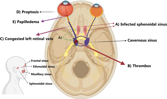
trauma, smoking, oral contraceptive pills use, or previous surgery (Figure 2).2,5 The aforementioned risk factors were not declared by the patient during the clinical
interview. This supports the hypothesis of a possible inherited PCD triggered by an
unidentified infection.4 There have been some cases reported of patients without any family history of hypercoagulable
states that developed a thrombotic event and found to have thrombophilia.2
Figure 2.
Simplified Graphic of an Infected Cavernous Sinus Thrombosis.

Legend: A) An infected sphenoid sinus causes septic thrombosis in the cavernous sinus, B) In cavernous thrombosis, the facial vein, and superior and inferior ophthalmic veins
C) cannot drain properly, resulting in facial and periorbital edema, ptosis, proptosis
D), chemosis, eye movement discomfort, papilledema (E), retinal vein dilation, and vision loss. This image was created with Biorender.
Due to undetermined timeline of sphenoidal rhinosinusitis, the recommended 10 days
antibiotic treatment was extended to 5 weeks by the infectious disease department.
According to the management of chronic rhinosinusitis described by Baron & Durand
in 2017, a minimum of 3 weeks of antibiotic course is recommended. Some symptoms of
chronic rhinosinusitis tend to reappear after 10 days of treatment.10
At 8 months, imaging studies were consistent with complete resolution of the thrombotic
event and sphenoidal rhinosinusitis. The continuation of anticoagulation with a direct
oral anticoagulant (Dabigatran) after hospitalization showed no recurrence of thrombotic
events, similar to what was reported by Fukushima et al. 9
The limitation of this case is the uncertainty about its infectious etiology. Cultures
resulted negative and a nasopharyngeal swab for bacteria was not performed. Due to
a possible chronic sphenoidal rhinosinusitis as the infectious source, empirical prolonged
antibiotic treatment was prescribed (this is debatable). Moreover, because of diplopia,
confrontation visual field examination could not be assessed correctly
Conclusions
Although rare, a patient without medical and family history of thrombophilia may develop
cerebral venous thrombosis because of Protein C Deficiency. Sinus infection may worsen
the clinical state. Early recognition with clinical examination and imaging studies,
followed by prompt intervention with anticoagulation and broad-spectrum antibiotics,
is associated with a good prognosis for patients with septic CST due to hypercoagulability.
Summary – Accelerating Translation
Este reporte de caso presenta a un paciente con trombofilia debido a una deficiencia
de proteina C (DPC) tipo 1 complicado por una trombosis del seno cavernoso (TSC).
La TSC séptica fue confirmada por celulitis periorbitaria y hallazgos de laboratorio.
El paciente también presentaba historia de dolores de cabeza de 3 meses, diplopía
y anormalidades fundoscópicas. La sospecha de DCP tipo 1 surgió debido a bajos niveles
plasmáticos de Proteína C asociados con un evento trombótico. La tomografía computarizada
mejorada con contraste y la resonancia magnética venográfica mostraron un aumento
y un defecto de llenado en el seno cavernoso izquierdo después de la administración
de contraste, así como tortuosidad y dilatación de las venas oftálmicas izquierdas.
Se recomendó un tratamiento prolongado de 5 semanas con antibióticos debido a una
posible rinosinusitis esfenoidal crónica como fuente infecciosa. En el seguimiento
a los 8 meses, se observó una resolución completa del evento trombótico y de la rinosinusitis
esfenoidal. Se realizó anticoagulación oral (Dabigatrán) después del alta hospitalaria,
lo que evitó la recurrencia de eventos trombóticos. Las limitaciones del caso incluyen
la falta de cultivos positivos y la ausencia de una muestra de hisopado nasofaríngeo
para bacterias.
Acknowledgments
We thank the excellent multidisciplinary team conformed by Dr. Nelson Maldonado, Dr.
Pablo de la Cerda, Dr. Catalina Salinas, and Dr. Marcos di Stefano, who contributed
to the management and resolution of this case. Finally, to this brave patient that
taught us the real significance of life.
Conflict of Interest Statement & Funding
The Authors have no funding, financial relationships or conflicts of interest to disclose.
Author Contributions
Conceptualization: WPS, and CBB. Validation: WPS. Formal Analysis: WPS, and CBB. Data
Curation: JDAE. Writing – Original Draft: WPS, and CBB. Writing – Review & Editing:
JDAE. Supervision: JDAE.
References
1. Martinelli I, Passamonti SM, Bucciarelli P. Handbook of Clinical Neurology. 1st ed, Milan: Italy; 2014.
2. Majid Z, Tahir F, Ahmed J, Bin Arif T, Haq A. Protein C Deficiency as a Risk Factor for Stroke in Young Adults: A Review. Cureus. 2020;12(3).
3. Danese S, Vetrano S, Li Z, Poplis VA, Castellino FJ. The protein C pathway in tissue inflammation and injury: Pathogenic role and therapeutic
implications. Blood. 2010;115(6):1121–31.
4. Dinarvand P, Moser KA. Protein C deficiency. Arch Pathol Lab Med. 2019;143(10):1281–5.
5. Stam J. Thrombosis of cerebral veins and sinuses. N Engl J Med. 2005;352:1791–8.
6. Ferro JM, Canhão P, Stam J, Bousser MG, Barinagarrementeria F. Prognosis of Cerebral Vein and Dural Sinus Thrombosis: Results of the International
Study on Cerebral Vein and Dural Sinus Thrombosis (ISCVT). Stroke. 2004;35(3):664–70.
7. Bhatia H, Kaur R, Bedi R. MR imaging of cavernous sinus thrombosis. Eur J Radiol Open. 2020;(7):100226.
8. Weerasinghe D, Lueck CJ. Septic Cavernous Sinus Thrombosis: Case Report and Review of the Literature. Neuroophthalmology. 2016;40(6):263–76.
9. Fukushima T, Shimomura Y, Nagaya S, Morishita E, Kawakami O. A Case of Treatment With Dabigatran for Cerebral Venous Thrombosis Caused by Hereditary
Protein C Deficiency. Cureus. 2021;13(6):1–4.
10. Barshak MB, Durand ML. The role of infection and antibiotics in chronic rhinosinusitis. Vol. 2, Laryngoscope
Investig. Otolaryngol. 2017;(1):36–42.
Wilson S. Peñafiel-Pallares, 1 School of Medicine, Universidad de las Américas, Quito, Ecuador.
Camila Brito-Balanzátegui, 1 School of Medicine, Universidad de las Américas, Quito, Ecuador.
Jaime David Acosta-España, 2 Institute of Microbiology, Friedrich Schiller University Jena, Jena, Germany.
3 Senior Lecturer, Postgraduate Program in Infectious Diseases, School of Medicine, Pontificia Universidad Católica del Ecuador, Quito, Ecuador.
4 Centro de Investigación para la Salud en América Latina (CISeAL), Pontificia Universidad Católica del Ecuador, Quito, Ecuador.
About the Author: WSP is a fifth-year (5/6) medical student from Quito-Ecuador. He is interested in
anesthesiology, pediatrics, and internal medicine. His focus is on clinical practice
and research. He has experience in public and private health centers in the country
and abroad. His volunteer work is broad and focuses mainly on providing health services
to the neediest communities in South America.
Correspondence: Jaime David Acosta-España. Address: Schloßgasse 10, 07743 Jena, Germany. Email: jdae_14@hotmail.com
Editor: Francisco J. Bonilla-Escobar
Student Editors: Purva Shah, Surobhi Chatterjee & Benjamin Liu
Proofreader: Laeeqa Manji
Layout Editor:Ana Maria Morales
Process: Peer-reviewed
Cite as
Peñafiel-Pallares WS, Brito-Balanzátegui C, Acosta-España JD. Hypercoagulability and
Cavernous Sinus Thrombosis due to Protein C Deficiency. A Case Report. Int J Med Stud.
2023 Jan-Mar;10(1):76-79.
Copyright © 2023 Wilson S. Peñafiel-Pallares, Camila Brito-Balanzátegui, Jaime David
Acosta-España.
This work is licensed under a Creative Commons Attribution 4.0 International License.
International Journal of Medical Students, VOLUME 11, NUMBER 1, March 2023

