Case Report
Aberrant Right Subclavian Artery: Cadaver Case Report
Jason Kopp1, Ahmad Irshaid1, Justin Baker2, John Fitzsimmons3, Judith C. Lin4
doi: http://dx.doi.org/10.5195/ijms.2022.1647
Volume 10, Number 4: 413-416
Received 09 09 2022;
Rev-request 30 09 2022;
Rev-recd 17 11 2022;
Accepted 30 11 2022
ABSTRACT
Different aortic arch branching patterns exist in the general population. These branching
patterns can be benign or can cause a variety of symptoms in patients. In the case
of a more benign branching pattern, anomalies often go undiagnosed until discovered
postmortem. Case: While examining the anatomy of a cadaver in a medical school gross
anatomy course, an aortic arch anomaly was discovered. This variant is consistent
with an aberrant right subclavian artery (ARSA). In this variant, the right subclavian
artery branches from the most distal part of the aortic arch and runs both retrotracheal
and retroesophageal as it courses to the right shoulder. This variant is a result
of aberrant development of the aortic arch and may present with symptoms such as dysphagia
and shortness of breath, if any at all. In addition to the ARSA, there exist a common
bicarotid trunk and a direct branching of the left vertebral artery from the aortic
arch, both of which are rare anomalies. The cadaver’s medical history includes dysphagia
and stretched esophagus, although the severity is unknown. Conclusion: this case report
draws attention to these rare anatomical anomalies and includes a discussion of the
most common clinical presentation, and surgical implications of an aberrant right
subclavian artery anomaly
Keywords:
Aberrant Right Subclavian Artery;
Case Reports;
Aortic Arch;
Dysphagia (Source: MeSH-NLM).
Highlights:
- A cadaver case was discovered to have an aberrant right subclavian artery.
- There are many variants in the branching patterns of the aortic arch and highlighted
here is one of those specific patterns.
- Disruption in the embryonic development of the aortic arch leads to a wide variety
of branching patterns in the general population.
- Most patients are asymptomatic, but dysphasia lusoria is the most common presenting
symptom.
Introduction
The aortic arch is a critical structure in the cardiovascular system, as it is the
beginning of the systemic arterial circulatory system. Formation of the aortic arch
begins during the fourth week of embryonic development and is ultimately derived from
multiple structures. At this time, a structure called the aortic sac starts to develop.
During the fifth week of development, the aortic sac begins to grow and branch off
into the two dorsal aortas and the ventral aorta. Six paired pharyngeal aortic arches
develop, which connect the ventral and dorsal aortae.1 Some of these arches completely regress while portions of the other persist as the
mature aorta develops.1 The primary origins of the aortic arch, from proximal to distal, are the aortic sac,
the left fourth aortic arch, and the dorsal aorta.2
An aberrant right subclavian artery (ARSA) affects approximately 1% of the population.2 ARSA develops as a result of the abnormal regression of the right fourth aortic arch
and right dorsal aorta distal to the right common carotid artery.1 In this case, the aberrant right subclavian develops from the right seventh segmental
artery of the descending aorta and becomes the most distal branch of the aortic arch.1,2 The aberrant right subclavian artery passes retrotracheal and retroesophageal in
80% of the cases.2 Alternatively, it courses between the trachea and the esophagus in 15% of the cases,
and anterior to the trachea in 5% of the cases.2 In addition to an aberrant right subclavian artery, there can also be the occurrence
of a bicarotid trunk. The coexistence of both ARSA and the bicarotid trunk has an
estimated prevalence of <0.05%.3
A majority of individuals with an aberrant right subclavian artery are asymptomatic,4 and it is estimated that 60-80% of individuals with this anomaly will never develop
any symptoms.4 For those who do develop symptoms, it is more common for them to have dysphagia,
caused by mechanical compression of the esophagus by the aberrant artery. This is
referred to as dysphagia lusoria.5 Dysphagia lusoria is seen in 71.2% of symptomatic individuals, but can range from
only mild intermittent dysphagia to a potentially severe, and continuous dysphagia
of both solids and liquids.6 These symptoms usually appear in the middle to older age groups. However, the exact
reason for this is unclear.5 Other symptoms and their prevalence in symptomatic individuals include dyspnea (18.7%),
retrosternal pain (17.0%), cough (7.6%), and weight loss (5.9%).5 This case report highlights a patient with a right aberrant subclavian artery and
a bicarotid trunk who experienced dysphagia. Uniquely, our report includes gross anatomical
imaging and discusses the clinical implications of such an anomaly.
The Case
While examining cadavers as part of a medical school gross anatomy course, an anomalous
origin of the right subclavian was discovered. The variant was consistent with the
aberrant right subclavian artery, as previously described. In this case, the right
subclavian artery originates from the aortic isthmus rather than the brachiocephalic
trunk.
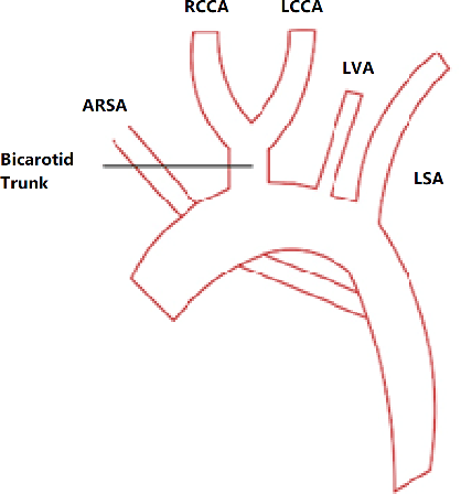
Figure 1 depicts the presentation of the aortic arch and its branches in our cadaver. The
left subclavian artery and left vertebral artery branch separately travel to the left
shoulder and head, respectively. The right and left common carotid arteries arise
from a common bicarotid trunk prior to coursing up the lateral portion of each side
of the neck.
Figure 1.
Schematic Depiction of the Aortic Arch and its Branches in the Cadaver.

Legend: ARSA = Aberrant Right Subclavian Artery; LCCA = Left Common Carotid Artery; LSA =
Left Subclavian Artery; LVA = Left Vertebral Artery; RCCA = Right Common Carotid Artery.
Figure 2 are photographs taken of the cadaver’s ARSA. The cadaver’s ARSA courses are posterior
to the trachea and esophagus. This is the most common ARSA variant, occurring in nearly
80% of cases.7
Figure 2.
Schematic Depiction of the Aortic Arch and its Branches in the Cadaver.

Legend: Left: Overhead View of the Cadaver Shows the Aberrant Origin of the Right Subclavian
Artery. The Proximal Aortic Arch is Being Reflected to the Left in Order to Display
the Very Posterior Origin. Right: A Lateral View of the Right Side of the Cadaver
Shows the Right Subclavian Artery Exiting Retrotracheal and Retroesophageal.
Dissection of the cadaver was performed as part of a prosection course at the Michigan
State University College of Human Medicine. The cadaver was dissected to reveal neurovascular
structures of the mediastinum and deep thorax, to include the subclavian vessels.
Our patient is an 81 year old male of unknown ethnicity with a past medical history
of aspiration pneumonia, diabetes, and Alzheimer’s disease. The causes of death noted
for the cadaver are cardiac arrest, acute respiratory failure, acute kidney injury,
and congestive heart failure. In addition to many other comorbidities, additional
medical information includes a history of stretched esophagus and dysphagia. Although
the severity of these medical conditions is unknown in the cadaver’s history, dysphagia
is often one of the sole symptomatic indicators of an ARSA.
Discussion
During a routine examination of cadavers as part of a gross anatomy course, an aberrant
right subclavian artery was discovered. This specific aortic arch anomaly is only
present in about 1% of people.2 However, in addition to the aberrant right subclavian artery, there was also the
occurrence of a bicarotid trunk, which is an extremely rare variant. The coexistence
of both ARSA and the bicarotid has an estimated prevalence of <0.05%.3 Lastly, the left vertebral artery arises from the aortic arch, which is estimated
to occur in around 4% of all individuals. Nevertheless, the combination of all three
has not been well documented.8 Therefore, the incidence cannot be determined in this report.
ARSA variations have been reported in the literature and Adachi?Williams have classified
them into three types based on the branches coming off of the aortic arch. Type I
(Type-G) has four branches: the right common carotid artery (RCCA), left common carotid
artery (LCCA), left subclavian artery (LSA), and aberrant right subclavian artery
(ARSA). Type II (Type CG) is the same as type I along with the addition of a branch
for left vertebral artery (LVA). Lastly, type III (type H) is seen with a bicarotid
trunk, LSA, and ARSA branches coming from the aorta.9 The case seen here is a combination of type II and type III variations due to the
presence of four branches: the LVA (seen in type II) and the bicarotid trunk (seen
in type III), along with LSA, and ARSA.
A recent systematic review, aimed at categorizing left sided aortic arch variants,
classified the variants based on the number of emerging branches into type 1b (one
branch), 2b, 3b, 4b, 5b and further subclassified based on prevalence.10 The systemic
review concluded that a typical branching pattern had a prevalence of 78% with a 22%
prevalence for all other branching patterns. The most common variant was 2b, with
bovine arch (LCCA originating from the brachiocephalic arch) being the most common
subtype of the 2b variant.
This case report is especially useful for examining an aortic arch anomaly and uniquely
presents gross anatomical findings as evidence of the anomaly. This report also includes
rare evidence of an ARSA coexisting with a bicarotid trunk. An associated history
of dysphagia in the cadaver helps demonstrate possible clinical findings of a patient
with an aortic arch anomaly. Due to the nature of the case report, we were unable
to examine the patient while they were still living and experiencing symptoms. The
findings of the case report were limited to the patient’s brief medical history reported
to the anatomy lab. Other cadaver case reports have reported similar findings of an
ARSA, and included more information on the clinical severity of the patient’s anomaly.11,12 However, future cadaver case reports should include thorough and complete clinical
descriptions of patients’ dysphagia as well as record of physician assessments and
surgical recommendations made during the clinical course. Diagnosing an ARSA in a
living patient is rare and is usually accidently discovered on a coronary angiogram.
Surgery may be implicated in extreme cases of dysphagia but is usually foregone in
favor of supportive treatment due to the risks of the procedure.
Individuals with ARSA variations can be asymptomatic and those with symptoms often
do not present until later in life, which makes a clinical diagnosis challenging.
Despite this, it is important that physicians consider an ARSA anomaly when a patient
presents with dysphagia, shortness of breath, and/or stridor. Other concerning symptoms
for a suspected ARSA include worsening retrosternal pain, cough, and acute limb ischemia.5 Although a surgical intervention is unlikely except for in rare and severe cases,
this case report raises clinical awareness of the possibility of such a condition
and can guide more specific intervention and treatment.
Summary – Accelerating Translation
The title of this case report is Aberrant Right Subclavian Artery: Cadaver Case Report.
The aorta is the major blood vessel leaving the heart and provides oxygenated blood
to the tissues. The aortic arch gives off 3 branches in 78% of the population. Normal
and pathological variation in the branching patterns and in the number of branches
exists in the general population. This case presentation discusses an aberrant right
subclavian artery (ARSA) in a cadaver. In the normal variant, the right subclavian
artery branches off the brachiocephalic artery, the most proximal branch of the aortic
arch. In our case, the right subclavian artery is the most distal branch of the aortic
arch and branches directly off of it as it courses behind the trachea and esophagus
to the right shoulder.
Most patients with this anomaly are generally asymptomatic, and the few that develop
symptoms tend to develop them later in life. Trouble swallowing, also called dysphagia
lusoria, is the most common symptom associated with ARSA. The abnormal position and
path of the right subclavian artery mechanically compresses the esophagus causing
the dysphagia. Other less common symptoms include shortness of breath, chest pain,
cough, and weight loss. Although interventions are generally rare, it’s important
for physicians to consider ARSA when working up a patient with dysphagia, and to be
aware of the variation in the branching patterns of the aortic arch when planning
for cardiothoracic interventions.
Acknowledgments
None.
Conflict of Interest Statement & Funding
The Authors have no funding, financial relationships or conflicts of interest to disclose.
Author Contributions
Conceptualization: JK, AI, JB, JF, JCL; Data Curation: JF; Formal Analysis: JK, AI,
JB, JF, JCL; Investigation: JK, AI, JB, JF; Project Administration: JF, JCL; Supervision:
JF, JCL; Writing - Original Draft Preparation: JK, AI, JB; Writing - Review & Editing:
JK, AI, JB, JF, JCL.
References
1. Hanneman K, Newman B, Chan F. Congenital Variants and Anomalies of the Aortic Arch. RadioGraphics. 2017;37(1):32–51. doi: http://dx.doi.org/10.1148/rg.2017160033

2. Rosen RD, Bordoni B. Embryology, Aortic Arch. National Library of Medicine NCBI. https://www.ncbi.nlm.nih.gov/books/NBK553173/. Published February 10, 2022.
3. Hanžič N, Čizmarević U, Lesjak V, Caf P. Aberrant right subclavian artery with a bicarotid trunk: The importance of diagnosing
this rare incidental anomaly. Cureus. 2019. doi: http://dx.doi.org/10.7759/cureus.6094

4. Brauner E, Lapidot M, Kremer R, Best LA, Kluger Y. Aberrant right subclavian artery-suggested mechanism for esophageal foreign body impaction:
Case report. World Journal of Emergency Surgery. 2011;6(1):1–3. doi: http://dx.doi.org/10.1186/1749-7922-6-12

5. Polguj M, Chrzanowski Ł, Kasprzak JD, Stefańczyk L, Topol M, Majos A. The aberrant right subclavian artery (Arteria Lusoria): The morphological and clinical
aspects of one of the most important variations—A systematic study of 141 reports. The Scientific World Journal. 2014;2014:1–6. doi: http://dx.doi.org/10.1155/2014/292734

6. Irakleidis F, Kyriakides J, Baker D. Aberrant right subclavian artery – a rare congenital anatomical variation causing
dysphagia Lusoria. Vasa. 2020;50(5):394–397. doi: http://dx.doi.org/10.1024/0301-1526/a000904

7. Natsis K, Didagelos M, Gkiouliava A, Lazaridis N, Vyzas V, Piagkou M. The aberrant right subclavian artery: Cadaveric Study and Literature Review. Surgical and Radiologic Anatomy. 2016;39(5):559–565. doi: http://dx.doi.org/10.1007/s00276-016-1796-5

8. Onrat E, Uluışık IE, Ortug G. The left vertebral artery arising directly from the aortic arch. Translational Research in Anatomy. 2021;24:100122. doi: http://dx.doi.org/10.1016/j.tria.2021.100122

9. Ramesh Babu CS, Gupta OP, Kumar A. Aberrant right subclavian artery: A multi-detector computed tomography study. Journal of the Anatomical Society of India. 2021;70(1):11–18. doi: http://dx.doi.org/10.4103/jasi.jasi_129_20

10. Natsis K, Piagkou M, Lazaridis N, et al. A systematic classification of the left-sided aortic arch variants based on cadaveric
studies’ prevalence. Surg Radiol Anat. 2021;43(3):327–345. doi: http://dx.doi.org/10.1007/s00276-020-02625-1

11. Abdelazeem B, Qureshi M, Alnaimat S, et al. Anomalous right subclavian artery as cause of dysphagia. BMJ Case Reports CP 2022;15:e247227.
12. Alghamdi MA, Al-Eitan LN, Elsy B, et al. Aberrant right subclavian artery in a cadaver: a case report of an aortic arch anomaly. Folia Morphol (Warsz). 2021;80(3):726–729. doi: http://dx.doi.org/10.5603/FM.a2020.0081
 .
.
Jason Kopp, 1 Second-year Medical Student, Michigan State University College of Human Medicine,
East Lansing, United States of America
Ahmad Irshaid, 1 Second-year Medical Student, Michigan State University College of Human Medicine,
East Lansing, United States of America
Justin Baker, 2 B.S. Michigan State University, East Lansing, United States of America
John Fitzsimmons, 3 MD. Michigan State University College of Human Medicine, East Lansing, United States
of America
Judith C. Lin, 4 MD, MBA. Michigan State University Department of Surgery, East Lansing, United States
of America
About the Author: Jason Kopp is currently a second-year medical student at Michigan State University
College of Human Medicine in East Lansing, MI, USA. This program is a four-year program.
Correspondence: Jason Kopp. Address: 15 Michigan St NE, Grand Rapids, MI 49503, United States. Email:
koppjas1@msu.edu
Editor: Francisco J. Bonilla-Escobar;
Student Editors: Hang-Long (Ron) Li & Muhammad Romail Manan;
Proofreader: Laeeqa Manji;
Layout Editor: Ana Maria Morales;
Process: Peer-reviewed
Cite as
Kopp J, Irshaid A, Baker J, Fitzsimmons J, Lin JC. Aberrant Right Subclavian Artery:
Cadaver Case Report. Int J Med Stud. 2022 Oct-Dec;10(4):413-16.
Copyright © 2022 Jason Kopp, Ahmad Irshaid, Justin Baker, John Fitzsimmons, Judith
C. Lin
This work is licensed under a Creative Commons Attribution 4.0 International License.
International Journal of Medical Students, VOLUME 10, NUMBER 4, December 2022


